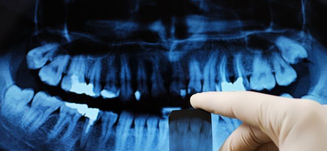
Getting routine dental X-rays is as important as brushing and flossing at home. Experts argue that having X-rays of a patient’s mouth is as important as the in-office dental cleaning. Why do you ask? Because a visual exam reveals half of the story: X-rays provide the other. Radiographs (a.k.a. X-rays) are a critical component of the dental exam. They provide essential information for tooth, gum, and bone health. Sometimes, a tooth can look normal to the eye but have a cavity lurking beneath the surface – only to be seen through an X-ray.
Furthermore, radiographs are very important to assess periodontal (gum and bone) health and changes in the bone that support the teeth. They are also important for the evaluation of oral pathology. Infections, cysts, or tumors may develop below the gums, or inside the bone where the dentist cannot see with the eye alone. Having radiograph information helps to accurately diagnose and treat dental problems early before they become more serious.
How Often Do X-rays Need to be Taken?
That answer depends on your medical and dental history. The American Dental Association recommends that new patients have posterior bitewings (molars and premolars) taken on their first visit. This helps the dentist determine the right treatment plan moving forward. During that first visit, the dentist may also recommend a new patient have panoramic images to assess overall health. This provides a baseline to help with future decisions
Generally speaking, if you have a great dental history, you can expect X-rays to be taken every 24 – 36 months. If you have a history of many cavities, fillings, or crowns, then X-rays are taken more often – possibly every six to 18 months.
What are the Different Types of X-Rays?
There are two main types of dental X-rays: intraoral and extraoral. The major difference in these two categories is that intraoral means that the X-ray film is placed inside your mouth and the opposite is true for extraoral – the film is placed outside of the mouth. Intraoral X-rays help to:
- Detect cavities
- Determine the health of the root of a tooth
- Provide information on bone health
- Check on developing teeth
Extraoral X-Rays help to detect health issues in the skull and jaw. Dentists use this category of X-rays to help diagnose and treat:
- Impacted teeth
- Tumors
- Cysts
- Fractures
- Problems in the gums, teeth, roots, and jaw
Intraoral X-rays are the most common type of X-ray and extraoral are often used by dentists placing implants or by orthodontists for teeth-realignment plans.
Is it Safe to Have X-Rays Taken?
Putting on that heavy, lead apron before a dental X-ray makes you wonder, is this safe? According to the American Dental Association, “Bitewing radiographs expose a patient to about 0.005 millisieverts (mSv) of radiation (a millisievert is a unit of measure). By comparison, because radiation is part of our environment, people in the United States are exposed, on average, to 3.2 mSv every year from background sources of radiation.”
In other words, we live in a radioactive world. From cell phones to microwaves, radiation surrounds us and is impossible to avoid. The low dose of radiation exposure in a dental office is small compared to other sources. Ultimately, the pros of having dental X-rays taken far outweigh the cons. They are proven to be an invaluable tool for treating dental health.